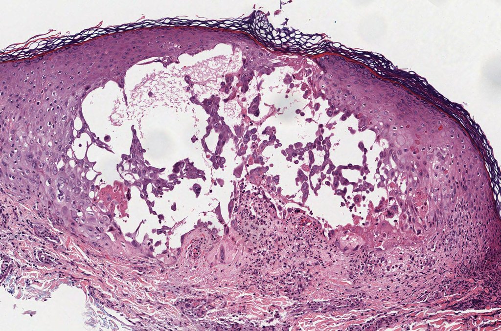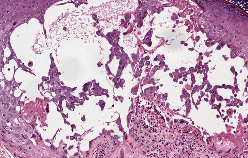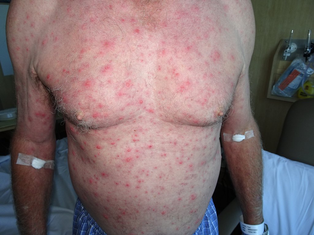
Diagnosis: Varicella
Description: Vesicle on chest wall
Clinical Features: Vesicles
Pathology/Site Features: Chest
Sex: M
Age: 25
Submitted By: Ian McColl
Differential DiagnosisHistory:
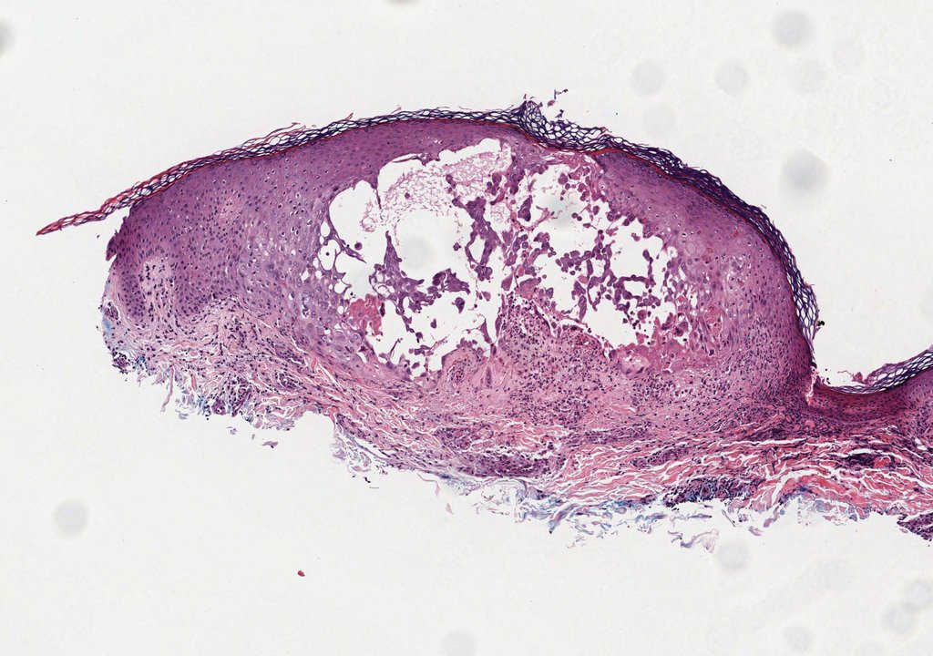
Histopathology of Herpes virus Dermnet on Herpes Simplex
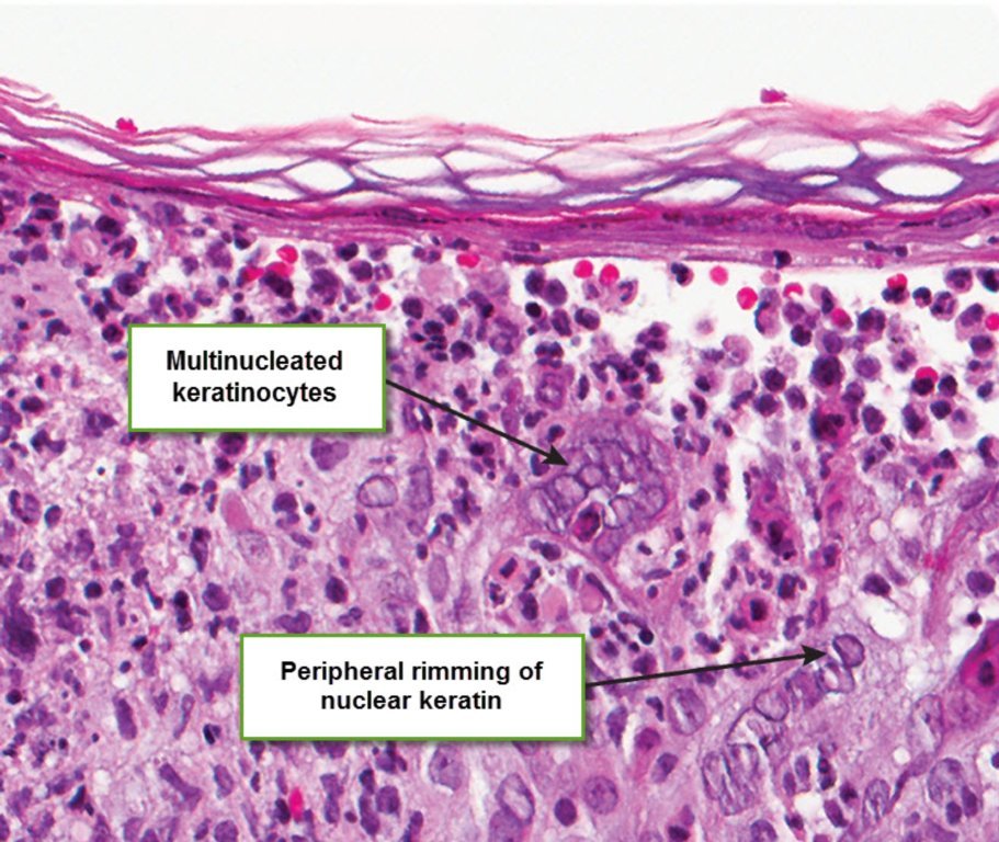
Clinical - Herpes simplex may be biopsied at the acute vesicular stage or at the later ulcerated stage particularly in immunosuppressed patients where healing is delayed. Histology of Herpes Simplex VS - Viruses cause both balloon and reticular degeneration of keratinocyes. Multinucleated keratinocytes and peripheral rimming of nuclear chromatin occur. Balloon degeneration is swelling of epidermal cells. Reticular degeneration with vesicles joining up and white septae is not specific for viral infections and can be seen in eczema. In some infected cells pink intranuclear inclusion bodies can be seen in some balloooned cells. Rarely atypical lymphoid infiltrates can be found under herpetic lesions. As herpes lesions age they may be infiltrated with neutrophils forming intraepidermal pustules. EM like pathology with basal vacuolar changes and increased numbers of apoptotic keratinocyes may also feature.There is also a lymphocytic vasculitis and sometimes a good marker is a perineural lymphocytic infiltrate.Vessels can also be involved with herpes virus infections. Differentials may include pemphigus vulgaris and Grovers in some cases with marked acantholysis. The changes in Varicella are very similar to herpes simplex with less inflammatory cells . Note also the verruciform variant in some HIV cases.
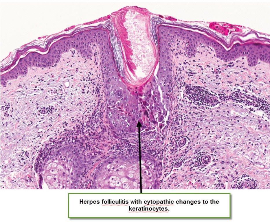
In follicular herpes simplex the same changes occur in the walls of the hair follicle.
